


Home / Albums / People / Physiology 110

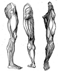 Muscles in the leg
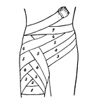 Ascending spica bandage of groin
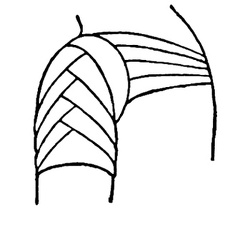 Ascending spica of shoulder
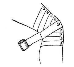 Bandage of the knee
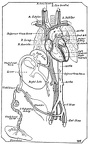 Diagrammatic view of the fetal circulation
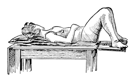 Dorsal recumbent posture
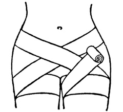 Double spica of groin
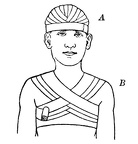 A, Recurrent bandage of the head - B, anterior figure-of-eight bandage of the chest
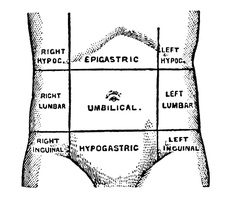 Abdominal regions
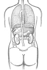 Position of the thoracic and abdominal organs, rear view
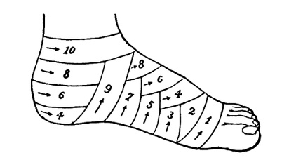 Spica bandage of ankle
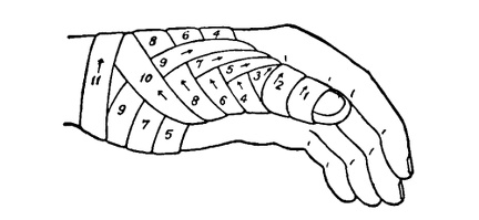 Spica bandage of thumb
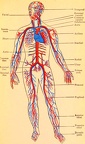 The principal arteries and veins of the body
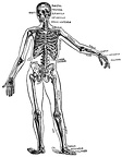 The skeleton
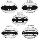 Eruption of the deciduous teeth
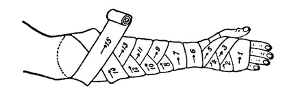 Figure-of-eight bandage of forearm
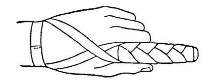 Finger bandage
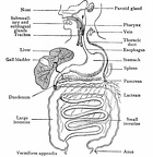 General scheme of the digestive tract
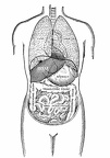 Position of the thoracic and abdominal organs, front view
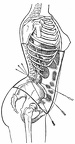 Diagram showing the action of the straight front corset
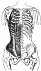 Muscles of the anterior surface of the trunk
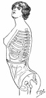 The abdominal corset
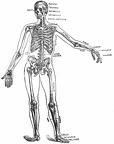 The skeleton
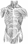 Muscles of the anterior surface of the trunk 2
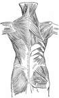 Muscles of the posterior surface of the trunk
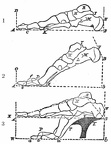 The natural and artificial positions of the foot
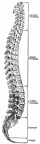 The spinal column
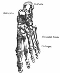 Upper surface, bones of foot
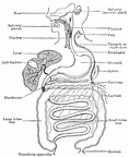 General scheme of the digestive tract
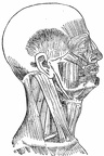 Muscles of the right side of the head and neck
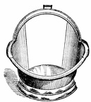 Sitz-bath tub made of tin
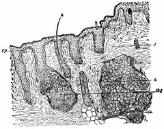 Vertical section of skin
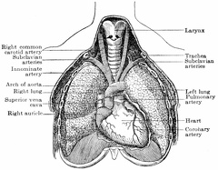 Front view of heart and lungs, showing relations to other thoracic organs
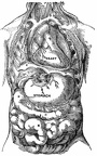 Location of the viscera of the body
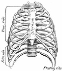 The bony thorax, anterior view
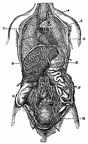 The ribs removed, showing relation of thoracic to abdominal viscera
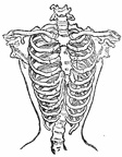 Effects of tight lacing on bony thorax
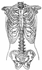 Normal chest
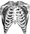 Relation of heart and great vessels to the wall of the thorax
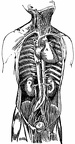 Relation of kidneys to heart and great blood-vessels
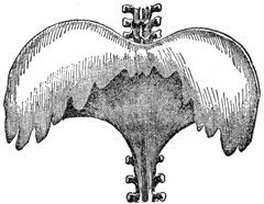 The diaphragm
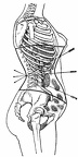 Diagram showing the action of the curved front corset
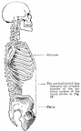 Skeleton of head and trunk
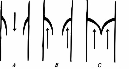 Diagram of Valves in the Heart and Veins
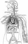 Diagram of the circulatory system
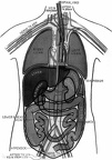 Outline diagram showing general plan and position of body-machinery
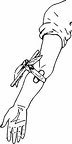 A Tourniquet
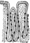 A longitudinal section of stomach, or peptic, glands
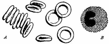 Blood Corpuscles
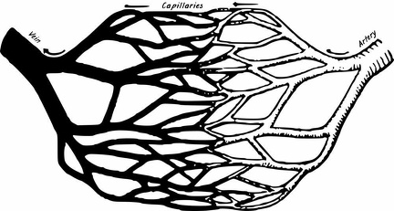 Diagram of artery, capillaries, and veins
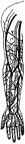 Surface veins and deep-lying arteries of inner side of right arm and hand
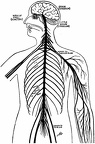 The Nervous System
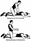 The New Method of Artificial Breathing
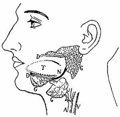 The Salivary Glands
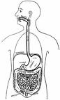 The food route in the digestive system
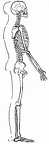 Human skeleton and Body outline
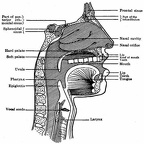 Section of the head and throat locating the organs of speech and song, including the upper resonators
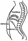 Figure shows the limit of full expiration and inspiration of the male, side view
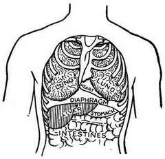 The diaphragm is in form like an inverted bowl
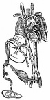 Plan of the foetal circulation