


Home / Albums / Natural History / Insects 209

 In the Honey Bee nearly all the mouth-parts of the Cockroach are to be made out, though some are sm...") Mouth-parts of Honey Bee
Mouth-parts of Honey Bee
In the Honey Bee nearly all the mouth-parts of the Cockroach are to be made out, though some are small and others extremely produced in length. The mandibles (Mn) are not much altered, and are still used for biting, as well as for kneading wax and other domestic work. The mandibular teeth have proved inconvenient, and are gone. The lacinia of the maxilla (Mx′) forms a broad and flexible blade, used for piercing succulent tissues, but the galea has disappeared, and there is only a vestige of the maxillary palp (Mxp). In the second pair of maxillæ the palp (Lp) is prominent; its base forms a blade, while the tip is still useful as an organ of touch. The paraglossæ (Pa) can be made out, but the laciniæ are fused to form the long, hairy tongue. This ends in a spoon-shaped lobe (not unlike the “finger” of an elephant’s trunk), which is used both for licking and for sucking honey. Chiasognathus Grantii, under side") Chiasognathus Grantii, under side
Chiasognathus Grantii, under side
Chiasognathus Grantii, under side") Chiasognathus Grantii 2
Chiasognathus Grantii 2 Chiasognathus Grantii, upper view") Chiasognathus Grantii, upper view
Chiasognathus Grantii, upper view
Chiasognathus Grantii, upper view 2 Chiasognathus Grantii, under side
3,4 Maxillae with lacinia and palpus
5 Mentumprocesses of labi...") Chiasognathus Grantii
Chiasognathus Grantii
2 Chiasognathus Grantii, under side 3,4 Maxillae with lacinia and palpus 5 Mentumprocesses of labium and palpi, under view 6 Base of anterior femora 7 Mentum, labium, &c. upper view 8 Labium with processes amd palpi, lateral view") Bees in Clover
Bees in Clover Cockroach-hunting wasps.
A, Dolichurus stantoni leading a nymph of Blattella lituricollis to her n...") Cockroach-hunting wasps
Cockroach-hunting wasps
Cockroach-hunting wasps. A, Dolichurus stantoni leading a nymph of Blattella lituricollis to her nest, c. X 4. B, Podium haematogastrum attaching her egg to an Epilampra sp. while on the side of a termite mound that contains the wasp's nest, c. X 1.6. C, Epilampra sp. parasitized by P. haematogastrum showing the wasp's egg attached to the right fore coxa, c. X 3.2. Protozoa from the gut of the wood-feeding cockroach Cryptocercus punctulatus.
A, Eucomonympha imla...") Protozoa from the gut of the wood-feeding cockroach
Protozoa from the gut of the wood-feeding cockroach
Protozoa from the gut of the wood-feeding cockroach Cryptocercus punctulatus. A, Eucomonympha imla, female above, male below, c. X 375. (From Cleveland [1950c].) B, Barbulanympha sp. (From Cleveland [1953].) C, Urinympha talea, c. X 712. (From Cleveland [1951a].) D, Rhynchonympha tarda, c. X 450. (From Cleveland [1952].) E, Trichonympha okolona or T. algoa, c. X 390. Diagram illustrating the relationship between a mature plant of Herpomyces stylopygae and the integu...") Relationship between a mature plant of Herpomyces stylopygae and the integument of Blatta orientalis
Relationship between a mature plant of Herpomyces stylopygae and the integument of Blatta orientalis
Diagram illustrating the relationship between a mature plant of Herpomyces stylopygae and the integument of Blatta orientalis. Richards and Smith have studied the life history of Herpomyces stylopygae on the oriental cockroach. The plants grow only on living cockroaches, and the infection is disseminated by contact.") Evania appendigaster
Evania appendigaster Representative Protozoa associated with cockroaches.
A, Monocercomonoides melolonthae, X 3094 (aft...") Representative Protozoa associated with cockroaches
Representative Protozoa associated with cockroaches
Representative Protozoa associated with cockroaches. A, Monocercomonoides melolonthae, X 3094 (after Grassé). B, Coelosporidium periplanetae, X 1310 (after Sprague); trophozoite with spores and chromatoid bodies. C, Endamoeba blattae, X 273 (after Kudo); trophozoite. D, Lophomonas striata, X 330 (after Kudo). E, Lophomonas blattarum, X 660 (after Kudo). F, Retortamonas blattae, X 3094 (after Wenrich). G, Nyctotherus ovalis, X 175 (after Kudo). H, Gregarina rhyparobiae, c. X 52: mature trophozoite attached to intestinal wall of Leucophaea maderae. (Redrawn from J. M. Watson [1945].) I, Diplocystis schneideri, c. X 14.4 (after Kunstler). J, Gregarina blattarum, c. X 57 (after Kudo). K, Protomagalhaesia serpentula, X 36 (after Pinto). L, Gamocystis tenax, magnification not known (after Schneider). The cockroach mite, Pimeliaphilus podapolipophagus") The cockroach mite
The cockroach mite
The cockroach mite, Pimeliaphilus podapolipophagus (Blatta orientalis). a, female; b, male; c, side view of female; d, young. After Marlatt, Entom. Bul...") Common Cockroach
Common Cockroach
(Blatta orientalis). a, female; b, male; c, side view of female; d, young. After Marlatt, Entom. Bull. 4, U.S. Dept. Agric. The young creature is hatched from the egg in a form closely resembling, on the whole, that of its parent, so that the term 'miniature adult' sometimes applied to it, is not inappropriate. The baby cockroach is known by its flattened body, rounded prothorax, and stiff, jointed tail-feelers or cercopods; the baby grasshopper by its strong, elongate hind-legs, adapted, like those of the adult, for vigorous leaping. showing proboscis formed by flexible maxillae (g) between the labial palps (p);c, face; e, eye; the...") A. Head of a typical Moth
A. Head of a typical Moth
showing proboscis formed by flexible maxillae (g) between the labial palps (p);c, face; e, eye; the structure m has been regarded as the vestige of a mandible. B. Basal part (b) of maxilla removed from head, with vestigial palp (p). Magnified. a, Diamond-back Moth (Plutella cruciferarum)
b, young caterpillar, dorsal view
c, full-grown cater...") Stages of the Diamond-back Moth
Stages of the Diamond-back Moth
a, Diamond-back Moth (Plutella cruciferarum) b, young caterpillar, dorsal view c, full-grown caterpillar, dorsal view d, side view e, pupa, ventral view. From Journ. Dept. Agric. Ireland, vol. I The simple nests and tubes that have been described are made by spiders, most of which spin no other...") Spiderweb
Spiderweb
The simple nests and tubes that have been described are made by spiders, most of which spin no other webs. The larger and better known cobwebs for catching insects are made by comparatively few species. On damp mornings in summer the grass-fields are seen to be half covered with flat webs, from an inch or two to a foot in diameter, which are considered by the weatherwise as signs of a fair day. These webs remain on the grass all the time, but only become visible from a distance when the dew settles on them. Figure is a diagram of one of these nests, supposed, for convenience, to be spun between pegs instead of grass. The flat part consists of strong threads from peg to peg, crossed by finer ones, which the spider spins with the long hind-spinnerets Long-legged, brown spiders, with two spinnerets longer than the others, and extending out behind the...") Agalenidæ
Agalenidæ
Long-legged, brown spiders, with two spinnerets longer than the others, and extending out behind the body. Figure is Agalena nævia, the common grass spider. They make flat webs, with a funnel-shaped tube at one side, in which the spider waits. A large family of spiders, varying greatly in shape, color, and habits. Most of them are dull colore...") Drassidæ
Drassidæ
A large family of spiders, varying greatly in shape, color, and habits. Most of them are dull colored, and live under stones, or in silk tubes on plants, and make no webs for catching insects. Their eyes are small, and arranged in two rows on the front of the head. Their feet have two claws and a bunch of flat hairs. The spinnerets are usually long enough to extend a little behind the abdomen. The figure is a Drassus, and the eyes as seen from in front. (415 visits)") Cockroach (Roach)
Cockroach (Roach)") Cicindela tuberculata - Larva
Cicindela tuberculata - Larva") Cicindela tuberculata
Cicindela tuberculata") Colymbetes rufimanus
Colymbetes rufimanus") Pterostichus opulentus - Larva
Pterostichus opulentus - Larva") Pterostichus opulentus
Pterostichus opulentus") Colymbetes rufimanus - Larva
Colymbetes rufimanus - Larva Dorcus punctulatus") Dorcus punctulatus
Dorcus punctulatus
Dorcus punctulatus Dryocora howittii - Larva") Dryocora howittii - Larva
Dryocora howittii - Larva
Dryocora howittii - Larva Dryocora howittii") Dryocora howittii
Dryocora howittii
Dryocora howittii Staphylinus oculatus") Staphylinus oculatus
Staphylinus oculatus
Staphylinus oculatus Stethaspis suturalis") Stethaspis suturalis
Stethaspis suturalis
Stethaspis suturalis Chætosoma scaritides") Chætosoma scaritides
Chætosoma scaritides
Chætosoma scaritides Stethaspis suturalis - Larva") Stethaspis suturalis - Larva
Stethaspis suturalis - Larva
Stethaspis suturalis - Larva Oral and digestive system of Deinacrida megacephala
1, mandibles
2, maxillæ
3, labrum
4, lab...") Oral and digestive system of Deinacrida megacephala
Oral and digestive system of Deinacrida megacephala
Oral and digestive system of Deinacrida megacephala 1, mandibles 2, maxillæ 3, labrum 4, labium 5, maxillary palpi 6, labial palpi 8, œsophagus 9, crop 10, gizzard 11, pancreas 12, stomach 13, biliary vessels 14, ilium 15, colon 16, anus. Body of an insect (Hymenoptera), showing the principal divisions
A, head
B, thorax
C, abdomen
a,...") Body of an insect
Body of an insect
Body of an insect (Hymenoptera), showing the principal divisions A, head B, thorax C, abdomen a, antenna c, compound eyes m, mandible s, simple eyes b, prothorax d, mesothorax k, metathorax 1W, fore-wing 2W, hind-wing n, coxa o, trochanter p, femur r, tibia t, tarsus 1 to 9 segments of the abdomen.") Insect Frame 4
Insect Frame 4") Butterfly and flower frame
Butterfly and flower frame") Butterfly frame
Butterfly frame") Insect Frame 2
Insect Frame 2") Insect Frame 3
Insect Frame 3") Insect frame
Insect frame Bird watching a butterfly") On the Watch
On the Watch
Bird watching a butterfly") Two birds watching a bug
Two birds watching a bug Birds chasing insects") The Pursuit
The Pursuit
Birds chasing insects A external view
B opened
C end view.
The eggs of the Cockroach are laid sixteen together in a ...") Egg-capsule of P. orientalis
Egg-capsule of P. orientalis
A external view B opened C end view. The eggs of the Cockroach are laid sixteen together in a large horny capsule. This capsule is oval, with roundish ends, and has a longitudinal serrated ridge, which is uppermost while in position within the body of the female. The capsule is formed by the secretion of a “colleterial” gland, poured out upon the inner surface of a chamber (vulva) into which the oviducts lead. The secretion is at first fluid and white, but hardens and turns brown on exposure to the air. In this way a sort of mould of the vulva is formed, which is hollow, and opens forwards towards the outlet of the common oviduct. Eggs are now passed one by one into the capsule; and as it becomes full, its length is gradually increased by fresh additions, while the first-formed portion begins to protrude from the body of the female. When sixteen eggs have descended, the capsule is closed in front, and after an interval of seven or eight days, is dropped in a warm and sheltered crevice. In Periplaneta orientalis it measures about ·45 in. by ·25 in.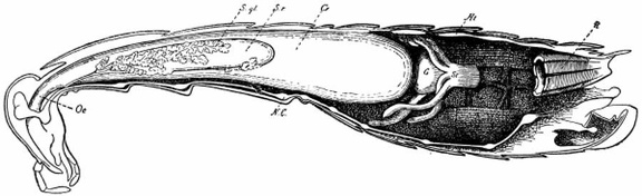 Longitudinal section of Female Cockroach
Longitudinal section of Female Cockroach, to show the position of the principal organs. Oe œsophagus S.gl, salivary gland S.r salivary reservoir Cr crop G gizzard St, chylific stomach R rectum Ht heart N.C nerve-cord Uses Of the uses to which Cockroaches have been put we have little to say. They constitute a popular remedy for dropsy in Russia, and both cockroach-tea and cockroach-pills are known in the medical practice of Philadelphia. Salted Cockroaches are said to have an agreeable flavour which is apparent in certain popular sauces. A female cockroach, Periplaneta, with the dorsal exoskeleton removed, dissected to show the viscera....") Cockroach
Cockroach
A female cockroach, Periplaneta, with the dorsal exoskeleton removed, dissected to show the viscera. 1 Head 2 labrum 3 antenna, cut short 4 eye 5 crop 6 nervous system of crop 7 gizzard 8 hepatic caeca 9 mid-gut or mesenteron 10 Malpighian tubules 11 colon 12 rectum 13 salivary glands 14 salivary receptacle 15 brain 16 ventral nerve cord with ganglia 17 ovary 18 spermatheca 19 oviduct 20 genital pouch, in which the egg-cocoon is found 21 colleterial glands 22 anal cercus (From Latter. Mouth appendages of Periplaneta (magnified).
A Mandible
B First maxilla
1 cardo
2 stipes
3...") Mouth appendages of cockroach
Mouth appendages of cockroach
Mouth appendages of Periplaneta (magnified). A Mandible B First maxilla 1 cardo 2 stipes 3 lacinia 4 galea 5 palp C Right and left second maxillae fused to form the labium 1 submentum 2 mentum 3 ligula, corresponding to the lacinia 4 paraglossa, corresponding to the galea 5 palp (From Latter.) Periplaneta orientalis, male.
Side view.
1 Antenna
2 head
3 prothorax
4 anterior wing
5 so...") Cockroach
Cockroach
Periplaneta orientalis, male. Side view. 1 Antenna 2 head 3 prothorax 4 anterior wing 5 soft skin between terga and sterna 6 sixth abdominal tergum 7 split portion of tenth abdominal tergum 8 cercianales 9 styles 10 coxa of third leg 11 trochanter 12 femur 13 tibia 14 tarsus 15,claws (From Kükenthal.) Periplaneta orientalis, male
Dorsal view.
1 Antenna
2 palp of first maxilla
3 prothorax
4 ante...") Cockroach
Cockroach
Periplaneta orientalis, male Dorsal view. 1 Antenna 2 palp of first maxilla 3 prothorax 4 anterior wings 5 femur of second leg 6 tibia 7 tarsus 8 cerci anales 9 styles (From Kükenthal.) Spirochætosis of Fowls—One of the best known of the spirochætes transmitted by arthropods is Spi...") Spirochæta gallinarum. After Hindle.
Spirochæta gallinarum. After Hindle.
Spirochætosis of Fowls—One of the best known of the spirochætes transmitted by arthropods is Spirochæta gallinarum, the cause of a very fatal disease of domestic fowls in widely separated regions of the world. According to Nuttall, it occurs in Southeastern Europe, Asia, Africa, South America and Australia. In 1903, Marchoux and Salimbeni, working in Brazil, made the first detailed study of the disease, and showed that the causative organism is transmitted from fowl to fowl by the tick Argas persicus. They found that the ticks remained infective for at least five months. Specimens which had fed upon diseased birds in Brazil were sent to Nuttall and he promptly confirmed the experiments. (a) Anterior part of venter
(b) second stage nymph
(c) capitulum
(d) dorsal aspect of female
(e)...") Ornithodoros moubata
Ornithodoros moubata
(a) Anterior part of venter (b) second stage nymph (c) capitulum (d) dorsal aspect of female (e) ventral aspect of female (f) ventral aspect of nymph (g) capitulum of nymph Ornithodoros moubata, the carrier of African relapsing fever, or "tick-fever," is widely distributed in tropical Africa, and occurs in great numbers in the huts of natives, in the dust, cracks and crevices of the dirt floors, or the walls. It feeds voraciously on man as well as upon birds and mammals. Like others of the Argasidæ, it resembles the bed-bug in its habit of feeding primarily at night. Dutton and Todd observed that the larval stage is undergone in the egg and that the first free stage is that of the octopod nymph. By trypanosomiasis is meant a condition of animal parasitism, common to man and the lower animals, i...") Trypanosoma brucei
Trypanosoma brucei
By trypanosomiasis is meant a condition of animal parasitism, common to man and the lower animals, in which trypanosomes, peculiar flagellate protozoa, infest the blood. Depending upon the species, they may be harmless, producing no appreciable ill-effect, or pathogenic, giving rise to conditions of disease. A number of these are known to be transferred by insects. The trypanosomes are elongated, usually pointed, flagellated protozoa in which the single flagellum, bent under the body, forms the outer limit of a delicate undulating membrane. It arises near one end of the organism from a minute centrosome-like body which is known as the blepheroplast, and at the opposite end extends for a greater or less distance as a free flagellum. Enclosing, or close beside the blepheroplast is the small kinetonucleus. The principal nucleus, round or oval in form, is situated near the center of the body. Asexual reproductions occurs in this stage, by longitudinal fission, the nucleus and the blepheroplast dividing independently of one another. From the blepheroplast of one of the daughter cells a new flagellum is formed. When the blood of an infested individual is sucked up and reaches the stomach of such a mosquito, th...") Filaria in the muscles and labium of Culex
Filaria in the muscles and labium of Culex
When the blood of an infested individual is sucked up and reaches the stomach of such a mosquito, the larvæ, by very active movements, escape from their sheaths and within a very few hours actively migrate to the body cavity of their new host and settle down primarily in the thoracic muscles. There in the course of sixteen to twenty days they undergo a metamorphosis of which the more conspicuous features are the formation of a mouth, an alimentary canal and a trilobed tail. At the same time there is an enormous increase in size, the larvæ which measured .3 mm. in the blood becoming 1.5 mm. in length. This developmental period may be somewhat shortened in some cases and on the other hand may be considerably extended. The controlling factor seems to be the one of temperature. The transformed larvæ then reenter the body cavity and finally the majority of them reach the interior of the labium. A few enter the legs and antennæ, and the abdomen, but these are wanderers which, it is possible, may likewise ultimately reach the labium, where they await the opportunity to enter their human host. Stomoxys calcitrans, the stable-fly—It is a popular belief that house-flies bite more viciously ju...") Stomoxys calcitrans - adult, larva, puparium and details
Stomoxys calcitrans - adult, larva, puparium and details
Stomoxys calcitrans, the stable-fly—It is a popular belief that house-flies bite more viciously just before a rain. As a matter of fact, the true house-flies never bite, for their mouth-parts are not fitted for piercing. The basis of the misconception is the fact that a true biting fly, Stomoxys calcitrans , closely resembling the house-fly, is frequently found in houses and may be driven in in greater numbers by muggy weather. From its usual habitat this fly is known as the "stable-fly" or, sometimes as the "biting house-fly." (577 visits) The house-fly breeds by preference in horse manure. Indeed, It has been found that they would develo...") The house or typhoid fly (Musca domestica)
The house or typhoid fly (Musca domestica)
The house-fly breeds by preference in horse manure. Indeed, It has been found that they would develop in almost any fermenting organic substance. Thus, they have been bred from pig, chicken, and cow manure, dirty waste paper, decaying vegetation, decaying meat, slaughter-house refuse, sawdust-sweepings, and many other sources. A fact which makes them especially dangerous as disease-carriers is that they breed readily in human excrement. - Caudal aspect - Anterior stigmata. Pharyngeal skeleton (563 visits) Sarcophagidæ—The larvæ of flies of this family usually feed upon meats, but have been found in c...") Larva of a flesh fly (Sarcophaga) - Caudal aspect - Anterior stigmata. Pharyngeal skeleton
Larva of a flesh fly (Sarcophaga) - Caudal aspect - Anterior stigmata. Pharyngeal skeleton
Sarcophagidæ—The larvæ of flies of this family usually feed upon meats, but have been found in cheese, oleomargerine, pickled herring, dead and living insects, cow dung and human feces. Certain species are parasitic in insects. Higgins (1890) reported an instance of "hundreds" of larvæ of Sarcophaga being vomited by a child eighteen months of age. There was no doubt as to their origin for they were voided while the physician was in the room. There are many other reports of their occurrence in the alimentary canal. We have recorded elsewhere (Riley, 1906) a case in which some ten or twelve larvæ of Sarcophaga were found feeding on the diseased tissues of a malignant tumor. The tumor, a melanotic sarcoma, was about the size of a small walnut, and located in the small of the back of an elderly lady. The chigoes, or true chiggers, are the most completely parasitic of any of the fleas. Of the dozen o...") Dermatophilus penetrans
Dermatophilus penetrans
The chigoes, or true chiggers, are the most completely parasitic of any of the fleas. Of the dozen or more known species, one commonly attacks man. This is Dermatophilus penetrans, more commonly known as Sarcopsylla penetrans or Pulex penetrans. This species occurs in Mexico, the West Indies, Central and South America. The males and the immature females of Dermatophilus penetrans closely resemble those of other fleas. They are very active little brown insects about 1-1.2 mm. in size, which live in the dust of native huts and stables, and in dry, sandy soil. In such places they often occur in enormous numbers and become a veritable plague.") Larva of Xenopsylla cheopis
Larva of Xenopsylla cheopis The whitish larvæ on hatching are slightly flattened ventrally, and each segment bears posteriorly ...") Larva of Auchmeromyia luteola
Larva of Auchmeromyia luteola
The whitish larvæ on hatching are slightly flattened ventrally, and each segment bears posteriorly three foot-pads transversely arranged. At night the larvæ find their way into the low beds or couches of the natives and suck their blood. Of the twenty or more species of this genus occurring in the United States the following are known t...") Culicoides guttipennis - mouth parts of adult
Culicoides guttipennis - mouth parts of adult
Of the twenty or more species of this genus occurring in the United States the following are known to bite: C. cinctus, C. guttipennis, C. sanguisuga, C. stellifer, C. variipennis, C. unicolor.