


") Bastard Gemsbok (Antilope leucophaea, Pallas
Bastard Gemsbok (Antilope leucophaea, Pallas") Bony skeleton of Hippopotamus
Bony skeleton of Hippopotamus![Waterbok (Antilope [Kobus] ellipsprymna, Ogilby)](_data/i/upload/2020/09/05/20200905194455-002aa0eb-sm.jpg "Waterbok (Antilope [Kobus] ellipsprymna, Ogilby) (781 visites)") Waterbok (Antilope [Kobus] ellipsprymna, Ogilby)
Waterbok (Antilope [Kobus] ellipsprymna, Ogilby), showing the dentition (860 visites)") Skull of Bear (Ursus), showing the dentition
Skull of Bear (Ursus), showing the dentition, Male, Central Africa (796 visites) White-Eared Antelope") White-Eared Antelope (A. leucotes), Male, Central Africa
White-Eared Antelope (A. leucotes), Male, Central Africa
White-Eared Antelope (882 visites)") Argali (Ovis Poli)
Argali (Ovis Poli)") Esquimaux Dog
Esquimaux Dog") Walrus, showing the upper incisors in the form of tusks
Walrus, showing the upper incisors in the form of tusks") The Mehari, or racing Camel
The Mehari, or racing Camel") Walrus skull, showing the powerful canine teeth
Walrus skull, showing the powerful canine teeth") Opossum
Opossum The Woolly Kangaroo") The Woolly Kangaroo
The Woolly Kangaroo
The Woolly Kangaroo") Head of Indian Elephant
Head of Indian Elephant (676 visites)") Manatee (Manatus Americanus)
Manatee (Manatus Americanus)") Halicore Dugong
Halicore Dugong") Dorset Ram
Dorset Ram") Cotswold
Cotswold") African Fat-Tailed Sheep
African Fat-Tailed Sheep") Sheep-washing in Australia
Sheep-washing in Australia") Sheep-shearing operations in Australia
Sheep-shearing operations in Australia") Two-year old Southdown sheep
Two-year old Southdown sheep") Rambouillet-Negretti Ram
Rambouillet-Negretti Ram") Negretti Merino Ram
Negretti Merino Ram") Lord Chesham's Shropshire
Lord Chesham's Shropshire") Flock of sheep in Australia, under a large Eucalyptus
Flock of sheep in Australia, under a large Eucalyptus The Devil") Devil
Devil
The Devil \" And he arose and went towards his father's house, but when he was still a great way off, his fathe...") The Return of the prodigal
The Return of the prodigal
" And he arose and went towards his father's house, but when he was still a great way off, his father saw him, and was sorry for him, and ran and embraced him. Then he told his father how he had sinned and had lost his title to be called the old man's son, but the father was so glad to have his son come back repentant, that he told his servants to bring the best clothing and a ring to put on his son. And he made a great feast, and they were merry, for he said, "This is my son that was as one dead to me and is now alive again; he was lost and is found." Jesus says it shall be so with all those who set their minds upon storing up riches in this world, r...") The Rich fool
The Rich fool
Jesus says it shall be so with all those who set their minds upon storing up riches in this world, rather than laying up treasures in heaven by pleasing God and working in His service. Death will come when they least expect it, and they will have to leave all their earthly riches, and go where no treasure has been laid up for them. What a foolish man the builder of the house shown in our picture must have been! Of course, when the...") The house built upon the sand
The house built upon the sand
What a foolish man the builder of the house shown in our picture must have been! Of course, when the wind blew and the waves dashed against his house, it would fall. Look how the sea has washed the foundation away, and how the roof is falling in! And the people; see how they are fleeing to save their lives! And all this calamity because he built his house upon the sand. But the other house, shown in the distance: how firmly that stands! What a bold front it offers to the waves, and how safely it resists the fury of the storm. Its foundations are sure, because they rest upon the solid rock. \" And they let down the net into the sea, but it enclosed so great a multitude of fishes that they c...") The wonderful draught of fishes
The wonderful draught of fishes
" And they let down the net into the sea, but it enclosed so great a multitude of fishes that they could not draw them up; and the net brake. Then Simon beckoned to his partners, James and John, who were in the other boat, that they should come and help them. And they came and filled both boats with the fishes, so that they began to sink. His parents were amazed when they saw Jesus in such company. But Mary, while she rejoiced at finding...") Son, why hast thou thus dealt with us
Son, why hast thou thus dealt with us
His parents were amazed when they saw Jesus in such company. But Mary, while she rejoiced at finding Him, gently said, "Son, why hast Thou thus dealt with us? Behold Thy father and I have sought Thee sorrowing." Jesus replied, "How is it that ye sought Me? Wist ye not that I must be about My Father's business?" When the angels had departed, the shepherds returned to Bethlehem; and there, in a stable, they foun...") The Shepherds worshipping the infant Jesus
The Shepherds worshipping the infant Jesus
When the angels had departed, the shepherds returned to Bethlehem; and there, in a stable, they found the infant Jesus, lying in a manger, watched over and cared for by His mother Mary and Joseph. And so great was the surprise and joy of the shepherds that they went out and told all they met of the wondrous things which they had seen. So King Herod first sent for the learned men of the Jews, the chief priests and scribes, and demande...") The Wise Men before the King
The Wise Men before the King
So King Herod first sent for the learned men of the Jews, the chief priests and scribes, and demanded of them where Christ should be born; and when they had replied that it was to be in Bethlehem, he secretly called the wise men before him, and inquired of them what time the star appeared. After getting the information he needed, he dismissed the wise men, bidding them to go to Bethlehem "and search diligently for the young child; and when ye have found Him," said Herod, "bring me word again, that I may come and worship Him also." The two vertical lines are exactly the same length—measure them and see. Short lines turned back a...") Optical Illusion in dress
Optical Illusion in dress
The two vertical lines are exactly the same length—measure them and see. Short lines turned back at either end make one seem short; extended lines make the other seem longer.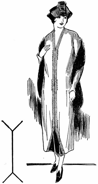
 These two illusions are almost duplicated in the dresses above. As a result one woman looks shorter ...") Optical Illusion in dress
Optical Illusion in dress
These two illusions are almost duplicated in the dresses above. As a result one woman looks shorter and heavier, the other taller and slenderer than she really is.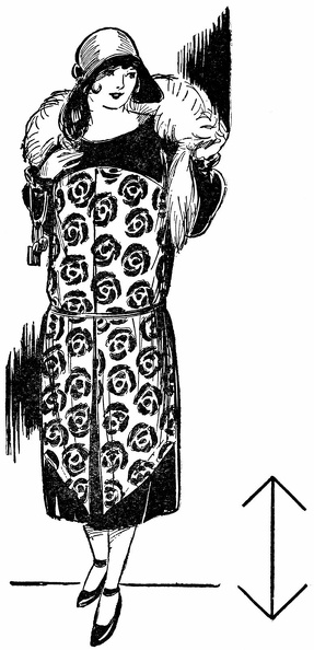
 These unbroken parallel vertical lines give the definite impression of height. This principle, used ...") Optical Illusion in dress
Optical Illusion in dress
These unbroken parallel vertical lines give the definite impression of height. This principle, used in the design of the dress above, lends it a pleasing slender appearance because no other lines interfere with the straight line effect.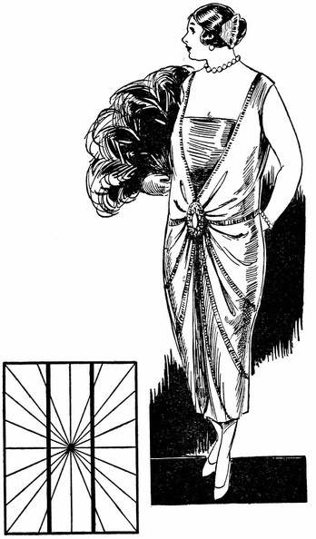
 Here, also, are two vertical parallel lines. They are straight—test them—but the other lines rad...") Optical Illusion in dress
Optical Illusion in dress
Here, also, are two vertical parallel lines. They are straight—test them—but the other lines radiating from the center, make them appear “bowed.” In the dress above a similar design makes the wearer appear stouter and heavier than she really is.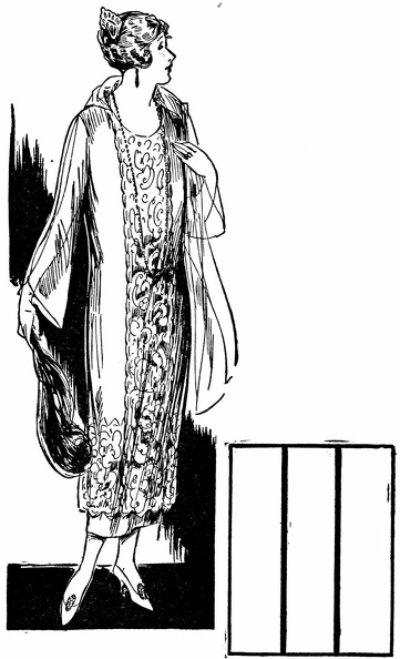
 These two diamond-shaped figures are exactly the same size. The crosswise line makes one seem wider,...") Optical Illusion in dress
Optical Illusion in dress
These two diamond-shaped figures are exactly the same size. The crosswise line makes one seem wider, the vertical line makes the other seem narrower.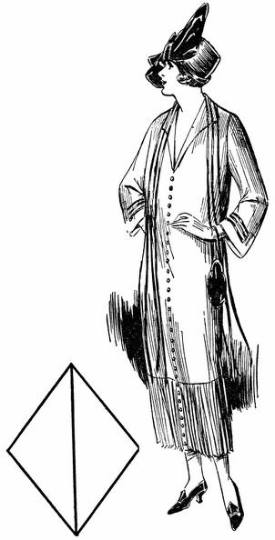
 Now note how these same principles used in the dresses above effect the apparent size and weight of ...") Optical Illusion in dress
Optical Illusion in dress
Now note how these same principles used in the dresses above effect the apparent size and weight of those wearing them, making one seem much stouter than the other.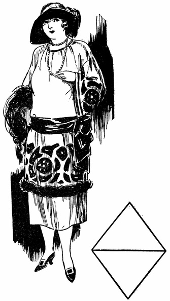
 The middle lines in the two small diagrams are the same length. But on the left, shorter accompanyin...") Optical Illusion in dress
Optical Illusion in dress
The middle lines in the two small diagrams are the same length. But on the left, shorter accompanying lines seem to shorten the one between. On the right longer accompanying lines seem to lengthen the one between.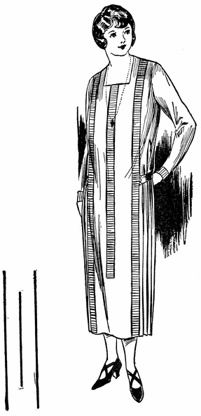
 Now see how the woman in the other picture has unknowingly emphasized her stoutness while the one in...") Optical Illusion in dress
Optical Illusion in dress
Now see how the woman in the other picture has unknowingly emphasized her stoutness while the one in this picure has properly gained a slender effect by using trimming in accordance with the principles of these optical illusions.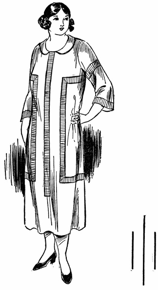
 The oblique line in the figure is made to seem longer and more graceful than the dress below by the...") Optical Illusion in dress
Optical Illusion in dress
The oblique line in the figure is made to seem longer and more graceful than the dress below by the parallel vertical lines of embroidery which intersect it and so emphasize its appearance of length and grace.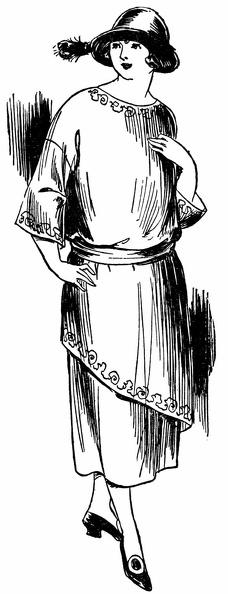
 When styles call for plaits, plaits may be used, but not in widening flares as shown here, rather in...") Plaits 1
Plaits 1
When styles call for plaits, plaits may be used, but not in widening flares as shown here, rather in slenderizing length lines as shown below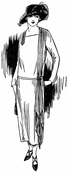
 Hats and shoes in these two pictures also illustrate incorrect and correct choice. The wide hat and ...") Plaits 2
Plaits 2
Hats and shoes in these two pictures also illustrate incorrect and correct choice. The wide hat and prominent straps below emphasize width and weight; the neat hat and cross-strap slippers here help to slenderize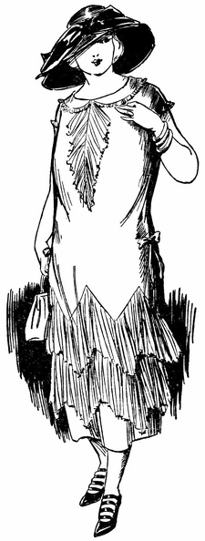
 These two pictures illustrate improper and proper choice of fabrics for a stout figure. Above, the l...") Choice of fabric 1
Choice of fabric 1
These two pictures illustrate improper and proper choice of fabrics for a stout figure. Above, the large-figured material adds size, the fur trim shortens, the round beads shorten the neck. All conspire to emphasize weight.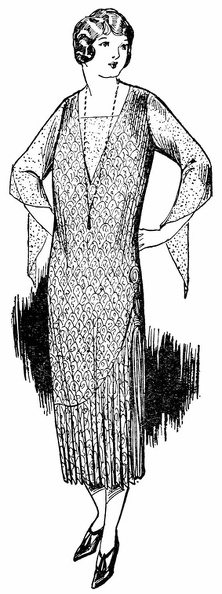
 Would you believe that the pattern of these two dresses is exactly the same? This illustrates how yo...") Two looks - same pattern
Two looks - same pattern
Would you believe that the pattern of these two dresses is exactly the same? This illustrates how you can vary a dress once you find the foundation lines that are becoming to you. One pattern can suffice for both a tailored and an afternoon dress, as you see both effects are pleasing in their slenderness. These two examples show how even a hat with drooping brim, if not too wide, can be worn by the stout...") Hats 1
Hats 1
These two examples show how even a hat with drooping brim, if not too wide, can be worn by the stout person if trimming is adeptly used to direct the vision upward and lend an illusion of height. Here trimming is used on two entirely different types of hats to give in each case added height to t...") Hats 2
Hats 2
Here trimming is used on two entirely different types of hats to give in each case added height to the figure and help in attaining a slenderizing appearance. Left—Hats with medium brims and high trimming are often becoming, especially if wide enough to avoid the pyramid effect. Right—High built trimming and delicate veils are advantageous where a double chin is the handicap. Here a small all-over pattern minimizes size, the plaits and tassels lengthen, the necklace adds a s...") Choice of fabric
Choice of fabric
Here a small all-over pattern minimizes size, the plaits and tassels lengthen, the necklace adds a slenderizing touch. The appearance as a whole is graceful and youthful. Note the diagonal line in the small diagram of the figure below. It is actually straight, but the ve...") Optical Illusion in dress
Optical Illusion in dress
Note the diagonal line in the small diagram of the figure below. It is actually straight, but the vertical lines which break it give it a “going-down-steps” appearance. This principle is used in the dress below—the two vertical panels of trimming break the line of the tunic and give the whole figure a more slender appearance than in the figure above.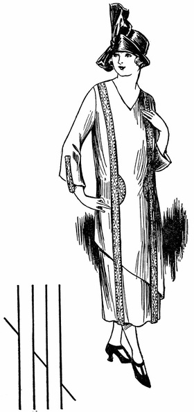
 Pear-shaped cells are set round a felt-work of nerve-fibrils (neuropil). A neuro-sensory cell is sho...") A Ganglion of a Leech
A Ganglion of a Leech
Pear-shaped cells are set round a felt-work of nerve-fibrils (neuropil). A neuro-sensory cell is shown with one fibre directed peripherally, branching on the surface; and one directed centrally, ramifying in the neuropil. Several very slender fibrils from the neuropil pass up the stalk of each ganglion-cell. They join a network near its surface. This net is connected by radiating fibrils with a coarser net which surrounds the nucleus. From the central net a relatively stout fibril passes to muscle-fibres. Half a dozen nuclei of as yet undeveloped granules are seen lying beneath the pia mater. From this l...") The Growth and Migration of Granules of the Cerebellum
The Growth and Migration of Granules of the Cerebellum
Half a dozen nuclei of as yet undeveloped granules are seen lying beneath the pia mater. From this level to the bottom of the drawing granules are shown in successive stages of growth. These developing granules, selected from various preparations of the cortex of the cerebellum, were drawn from nature. In its centre is a large clear spherical nucleus, with a nucleolus. The body-substance is prolonged ...") The Body of a Motor Neurone
The Body of a Motor Neurone
In its centre is a large clear spherical nucleus, with a nucleolus. The body-substance is prolonged into five dendrites and an axon. Neuro-fibrillæ are seen in dendrites and axon. They traverse the body of the cell in all directions, in little bundles which are separated by angular granules of stainable substance (tigroids). Sensory areas are enclosed by broken lines; certain centres in the association-zones are marked by d...") The Surface of the Left Cerebral Hemisphere, Cerebellum,and Medulla Oblongata.
The Surface of the Left Cerebral Hemisphere, Cerebellum,and Medulla Oblongata.
Sensory areas are enclosed by broken lines; certain centres in the association-zones are marked by dots. The sensory area of smell is on the inner aspect of the brain; so also is the area of vision which borders the calcarine and retrocalcarine fissures, and only rarely extends on to the external surface, as shown in the diagram. The sensory area of hearing is largely hidden within the fossa of Sylvius, the opening into which is indicated by the dark line above it. The kinæsthetic-sensory areas for the various muscles of the body occupy the territory between the dotted line in front and the bottom of the fissure of Rolando behind. They do not extend on to the posterior wall of this fissure. It is impossible at present to define the boundaries of any of the centres in the association-zones. These sense-organs are groups of elongated epithelial cells, set vertically to the surface. Their ce...") Highly Magnified Section through the Wall of a Circumvallate Papilla of the Tongue, showing Two Taste-Bulbs.
Highly Magnified Section through the Wall of a Circumvallate Papilla of the Tongue, showing Two Taste-Bulbs.
These sense-organs are groups of elongated epithelial cells, set vertically to the surface. Their cells are of two kinds—the one fusiform, slender, bearing each a bristle-like process which projects through a minute pore left between the superficial cells of the general epithelium; the other thicker and wedge-shaped. Nerve-fibres are connected with the fusiform cells. The slight depression in the retina in the axis of the globe is the fovea centralis, or yellow spot;...") Horizontal Section through the Right Eye
Horizontal Section through the Right Eye
The slight depression in the retina in the axis of the globe is the fovea centralis, or yellow spot; the optic nerve pierces the ball to its inner or nasal side. The lens, with its suspensory ligament, separates the aqueous from the vitreous humour. On the front of the lens rests the iris, covered on its posterior surface with black pigment. On either side of the lens is seen a ciliary process, with the circular fibres of the ciliary muscle cut transversely, and its radiating fibres disposed as a fan. A, after Exposure to Bright Light;
B, After Resting in the Dark.
The arrow shows the direction i...") The Retina in Vertical Section
The Retina in Vertical Section
A, after Exposure to Bright Light; B, After Resting in the Dark. The arrow shows the direction in which light traverses the retina. C, Retinal epithelium, with its pigmented fringe. 1, Layer of rods and cones, separated by the external limiting membrane from 2, the layer of the nuclei of the rods and cones. 3, The ganglion-cells of the retina, which are homologous with the cells of the afferent root of a spinal nerve. Their peripheral axons ramify beneath the sensory epithelium (rods and cones and their nucleus-bearing segments), their central axons in 4, the inner molecular layer. D, Collecting cells on the front of the retina; a a a, their axons which conduct impulses to the brain; b, an efferent fibre from the brain. x, The common centre of curvature (nodal point of the several media). Rays which pass through this p...") The Formation of an Image by the Refracting Media of the Eye
The Formation of an Image by the Refracting Media of the Eye
x, The common centre of curvature (nodal point of the several media). Rays which pass through this point are not deflected. y, The principal focus of the system. All rays which are parallel to the optic axis converge to this point. The image of the point A is formed at a, the spot at which a ray parallel with the optic axis meets an unbent ray—the image of B at b. From right to left, the figure shows the concha and lobule of the ear in profile; the external meatu...") The External, Middle, and Internal Ear of the Left Side
The External, Middle, and Internal Ear of the Left Side
From right to left, the figure shows the concha and lobule of the ear in profile; the external meatus (abbreviated); the drum, divided vertically, its posterior half visible; the hammer-bone, with the tip of its long arm attached to the drum, an arrow indicating the point of attachment and line of action of the tensor tympani muscle; the anvil attached by a ligament to the bony wall of the middle ear; the stirrup, with its foot-plate almost filling the oval window; the labyrinth, with the three semicircular canals above, and the scala vestibuli below. The curled black line shows the situation of the scala media, or ductus cochleæ (which contains the organ of Corti). Pulsations of sound which move the membrana tympani are transmitted by the three bones to the oval window. They shake the perilymph, producing waves which travel along the scala vestibuli to the apex of the cochlea, whence they return by the scala tympani to the round window (if they do not take a shorter course through the ductus cochleæ). The Eustachian tube opens out of the lower part of the middle ear. The spiral lamina, on the left of the drawing, gives attachment to the membrane of Corti, which stre...") Organ of Corti
Organ of Corti
The spiral lamina, on the left of the drawing, gives attachment to the membrane of Corti, which stretches to the opposite wall. Below the membrane is a bloodvessel which runs its whole length beneath the tunnel of Corti. The tunnel is formed by pillars—the inner on the left, the outer on the right—which meet above it. On the left of the inner pillar is a hair-cell; to the left of this a nerve-cell with two nuclei. To the right of the outer pillar is a space; to the right of this four hair-cells alternating with four supporting cells, which hold up the reticulated membrane through apertures in which the tufts of hairs project. Three nerve-fibres are seen in the spiral lamina; they cross the tunnel to ramify between the rows of outer hair-cells. The lamina tectoria rests upon the tufts of hairs.